
Introduction: The Invisible Universe
If you place two tiny dots on a piece of paper and move them closer and closer, there is a moment where they merge into one. At a distance of 25 cm—the “near point” of the human eye—our vision fails us if two objects are separated by less than 0.1 mm. This is our “limit of resolution.” It is a biological blind spot that keeps the most complex machinery in existence hidden from our view.
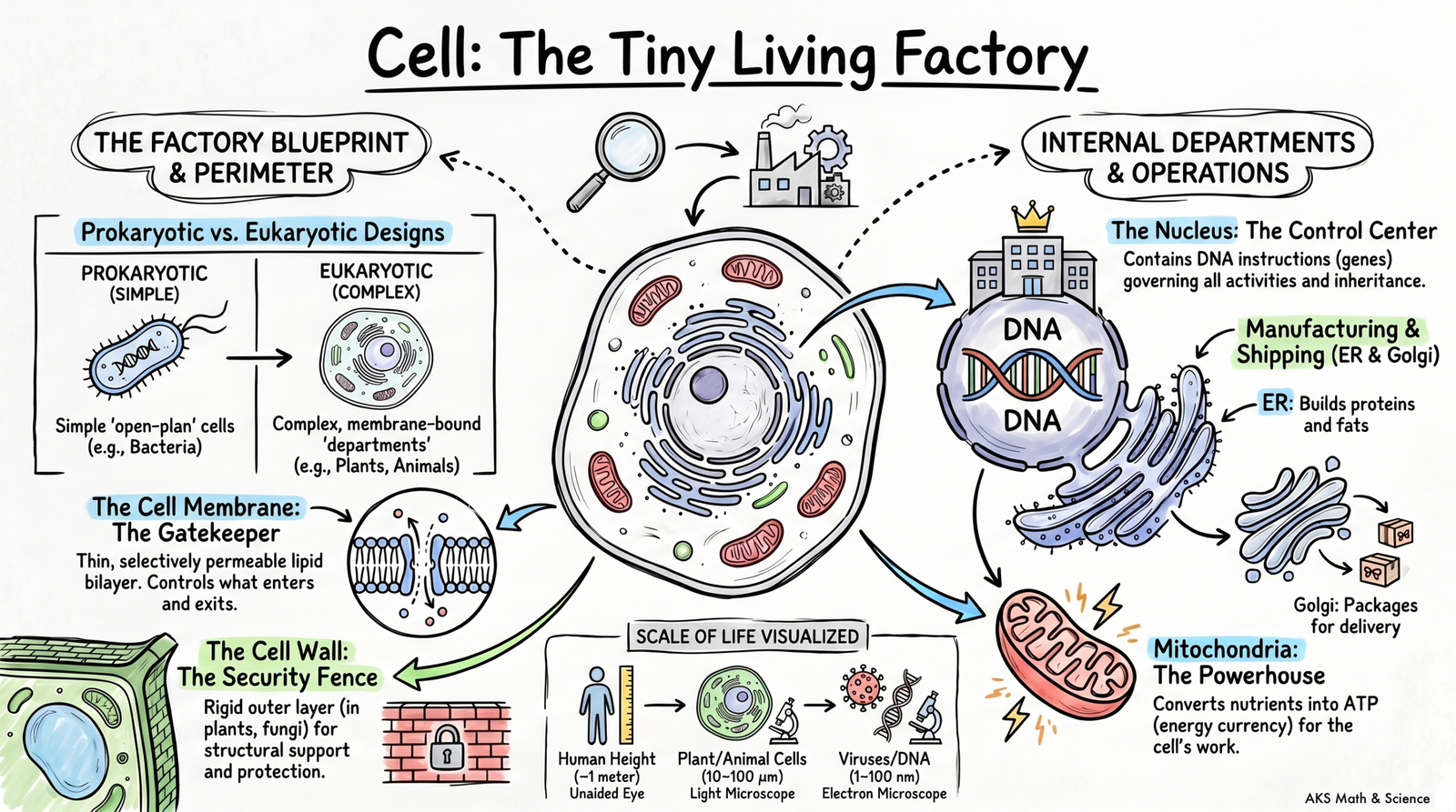
While we see ourselves as a single entity, we are actually vast “living factories” composed of trillions of individual units working in a coordinated system. By looking beyond this 0.1 mm threshold, we find that the cell is not just a building block; it is a sophisticated universe that redefines our understanding of life itself.
1. The Boiling Cradle: Life’s Unexpected Origin
We often imagine life began in the gentle shallows of a calm ocean. However, research from the Birbal Sahni Institute of Palaeosciences points to a far more violent birthplace: the near-boiling hot springs of Puga Valley in Ladakh. This extreme environment mirrors the harsh conditions of Earth 3.5 billion years ago.
In these springs, scientists discovered that calcium carbonate forms with incredible speed. This mineral didn’t just provide a roof; it acted as a “protective shield” that allowed life to make the jump from loose organic molecules to contained units. By safeguarding these molecules from lethal radiation, the mineral deposits likely midwifed the creation of the first cellular boundaries.
“The organisms living in these hot springs are mostly heat-loving bacteria called thermophiles… [Calcium carbonate] deposits may have protected early organic molecules from harmful radiation and extreme conditions, and they may have also helped in the formation of the first protective membrane — the barrier that defines a cell.”
2. The Fluid Gatekeeper: Why Your Boundaries Aren’t Solid
The plasma membrane is the universal boundary of life, but it is not a wall.
According to the “Fluid-Mosaic Model,” this 7 to 10 nanometre boundary is a shifting, dynamic sea.
It consists of a lipid bilayer where fat molecules rotate, move sideways, and flip.
This constant motion is critical for cellular communication and the life-sustaining flow of osmosis.
Embedded within this fluid sea are proteins that act as “gatekeepers.”
These proteins provide “selective permeability,” choosing exactly which molecules enter or exit.
The plant cell wall offers a stark contrast.
It is a rigid structure made of cellulose, designed to help the plant stay upright and withstand rain.
While the membrane is a picky bouncer, the cell wall is simply “permeable.”
It allows water and dissolved minerals to pass through relatively freely, relying on the membrane beneath it to do the actual sorting.
3. The Evolutionary Outsiders: Mitochondria and Plastids
Certain parts of your cells behave like biological foreigners. Mitochondria—the “powerhouses” that generate energy—and plastids, such as the food-synthesizing chloroplasts in plants, possess their own ribosomes and their own DNA.
This genetic independence is a smoking gun for an ancient evolutionary secret. These organelles were likely once independent, single-celled bacteria that were swallowed by larger cells billions of years ago. Instead of being digested, they stayed, forming a permanent partnership that changed the course of life.
“Mitochondria are often called the ‘powerhouses of the cell’ because they supply the energy needed for most cellular activities… Plants use special organelles called plastids for food synthesis and storage… these characteristics suggest that both mitochondria and plastids share an evolutionary history with these single-celled organisms.”
4. The Software of Life: Synthetic DNA and Cellular Control
To find the true “commander” of the cell, scientists performed a radical experiment: they replaced a bacterium’s biological soul. They stripped a host cell of its natural DNA, leaving behind only the “hardware”—the cytoplasm and the membrane.
Then, they inserted synthetic DNA into the empty shell. The result was transformative. The synthetic software completely hijacked the host’s hardware, forcing the cell to grow and divide according to the new, man-made instructions. This proved that while the cell is a complex machine, the DNA is the “coded software” that dictates every structural detail and every metabolic activity.
5. The Cost of a Copy: When Cell Division Goes Wrong
Cell division is a logistical feat of staggering proportions. Every single day, your body replaces roughly 1% of its total cell count—that is hundreds of billions of new units created every 24 hours.
This usually happens through two precise methods:
- Mitosis: The “controlled and orderly” production of identical daughter cells for growth and repair.
- Meiosis: The specialized division that creates genetic diversity for reproduction.
However, a system that makes 100 billion copies a day is statistically bound to face errors.
- Errors in Mitosis: When the “stop” signals fail, uncontrolled division creates tumors.
- Errors in Meiosis: Mistakes here lead to genetic disorders or reduced fertility.
The true surprise is not that these errors occur, but that they are so rare. Given the sheer mathematical improbability of rewriting the code for billions of cells daily without a total system collapse, our continued health is nothing short of a biological miracle.
Conclusion: The Quest Continues
The cell is far more than a “box-like compartment,” as Robert Hooke first described it in 1665. It is a highly coordinated working system that bridges the gap between chemistry and consciousness.
Today, we have moved beyond simple observation to “cell culture”—the ability to grow and manipulate these units in nutrient-rich mediums outside the body. As we learn to better script the “software” of life and sustain these “living factories” in the lab, we face a new frontier. If we can now control the very units that define us, what will the future of medicine and society look like? The quest to go beyond the 0.1 mm limit continues to redefine what it means to be alive.
FAQs
A cell operates like a tiny living factory because its organelles carry out various life processes independently yet simultaneously to maintain the cell’s survival and function. Each organelle has a specialized role, much like different departments in a manufacturing plant:
1. Management and Instructions (The Nucleus)
The nucleus acts as the “House of coded instructions”. It contains chromosomes made of DNA, which hold the genetic information (genes) required for the cell to function, grow, and divide. It directs all cellular activities, ensuring the “factory” follows the correct blueprints.
2. Manufacturing and Production (Ribosomes and ER)
- Ribosomes: These are the “protein factories” where protein synthesis actually occurs.
- Endoplasmic Reticulum (ER): Known as the “manufacturing factory,” the ER is a network that synthesizes and transports materials.
- The Rough ER (RER) has ribosomes attached and is primarily involved in making and secreting proteins.
- The Smooth ER (SER) lacks ribosomes and focuses on synthesizing and storing fats (lipids) and hormones.
3. Packaging and Distribution (Golgi Apparatus)
The Golgi apparatus serves as the cell’s “post office” or “packaging and shipping center”. It receives proteins and lipids from the ER, then modifies, sorts, and packages them into vesicles for transport to specific destinations or for secretion outside the cell.
4. Energy Supply (Mitochondria and Chloroplasts)
- Mitochondria: Called the “powerhouse of the cell,” they perform cellular respiration to break down molecules and release energy in the form of ATP (the cell’s energy currency).
- Chloroplasts (in plants): These are the “centers for food synthesis,” using sunlight and chlorophyll to produce food through photosynthesis.
5. Maintenance and Waste Management (Lysosomes)
Lysosomes function as the “clean-up system”. They contain enzymes that break down waste materials, damaged organelles, and foreign agents, keeping the cell clean and healthy.
6. Storage and Support (Vacuoles and Cytoskeleton)
- Vacuoles: These act as storage areas for water, minerals, and waste. In plant cells, a large central vacuole also provides structural pressure to keep the cell firm.
- Cytoskeleton: A network of fibers that provides the “factory” with its physical structure, maintains its shape, and enables internal transport.
By working together in this coordinated system, these organelles allow the cell to build new materials, remove waste, and generate the energy necessary for life.
The cell membrane (also called the plasma membrane) is considered the universal feature of life because every living cell is surrounded by one, regardless of whether it is a bacterium, a plant, or an animal.
According to the sources, the following characteristics make it fundamental to all life:
- Defining Individuality: The membrane acts as a thin boundary that protects the cell’s contents and defines its individuality as a distinct unit. It is the barrier that essentially “defines a cell”.
- Selective Permeability: It is selectively permeable, meaning it acts as a “gatekeeper” by allowing specific substances to pass through while blocking others. This allows the cell to maintain a stable internal environment through processes like osmosis (the diffusion of water) and the controlled movement of gases like oxygen and carbon dioxide.
- Communication and Interaction: All living cells must interact with their environment to survive. The membrane enables cells to communicate with their surroundings and neighboring cells, and it allows single-celled organisms to respond to environmental changes.
- Structural Composition: All cell membranes share a similar fluid-mosaic structure, consisting of a lipid bilayer (two layers of fat molecules) with embedded proteins that help transport materials.
- Evolutionary Significance: Scientific research suggests that the formation of the first protective membrane was a critical step in the origin of life approximately 3.5 billion years ago, as it protected early organic molecules from harsh external conditions.
While some organisms like plants, fungi, and bacteria have an additional outer layer called a cell wall for extra support, the cell membrane remains the primary, universal boundary for all living cells.
The fluid-mosaic model is the widely accepted scientific explanation for the structure of the cell membrane. It describes the membrane as an extremely thin boundary, approximately 7 to 10 nanometres thick, composed primarily of lipids (fats) and proteins.
According to the sources, the model is defined by the following characteristics:
- Lipid Bilayer: The foundation of the membrane is a double layer of special fat molecules called a lipid bilayer. These molecules have water-attracting heads facing outwards and water-repelling tails pointing inwards.
- The “Fluid” Nature: The membrane is considered fluid because the molecules within it are not fixed in place; they can move sideways, flip, and rotate.
- The “Mosaic” Nature: It is called a mosaic because the various molecules (lipids and proteins) are arranged together like tiles in a mosaic.
- Protein “Gatekeepers”: Various types of proteins are embedded throughout the lipid bilayer. These proteins act as gatekeepers, helping specific substances pass through the membrane to maintain the cell’s internal environment.
This structural arrangement allows the cell membrane to remain selectively permeable, controlling what enters and exits the cell while enabling communication with its surroundings.
Cells are categorized into two main types: prokaryotic (bacteria) and eukaryotic (plants and animals). While all three share certain features like a cell membrane and cytoplasm, they differ significantly in their internal organization and specialized structures.
1. The Nucleus and Genetic Material
- Bacterial Cells: These are prokaryotic, meaning they lack a well-defined nucleus. Their genetic material is not enclosed by a membrane and exists in a region called the nucleoid.
- Plant and Animal Cells: These are eukaryotic and contain a well-defined nucleus protected by a double-layered nuclear membrane.
2. Outer Boundaries
- Plant Cells: Have both a cell membrane and a rigid cell wall primarily made of cellulose, which provides structural support and protection.
- Bacterial Cells: Also possess a cell wall outside their cell membrane for protection.
- Animal Cells: Do not have a cell wall; they are surrounded only by a flexible cell membrane, allowing them to change shape easily.
3. Membrane-Bound Organelles
- Bacterial Cells: Lack membrane-bound organelles entirely; most cellular activities occur directly in the cytoplasm.
- Plant and Animal Cells: Contain various specialized membrane-bound organelles like mitochondria, the endoplasmic reticulum, and the Golgi apparatus.
4. Specialized Structures
- Plastids (Chloroplasts): These are found in plant cells to synthesize food through photosynthesis but are absent in animal and bacterial cells.
- Vacuoles: Plant cells typically have one large central vacuole that stores water and maintains cell firmness. Animal cells may have small, temporary vacuoles.
- Centrioles/Lysosomes: While the sources focus on plant and animal similarities, diagrams show lysosomes as a prominent feature in animal cells.
5. Size and Complexity
- Bacterial Cells: Generally much smaller, typically ranging from 1 to 10 µm in diameter.
- Plant and Animal Cells: Larger and more complex, typically ranging from 10 to 100 µm.
Comparison Summary Table
| Feature | Bacterial Cell | Plant Cell | Animal Cell |
|---|---|---|---|
| Type | Prokaryotic | Eukaryotic | Eukaryotic |
| Nucleus | Nucleoid (No membrane) | Well-defined | Well-defined |
| Cell Wall | Present | Present (Cellulose) | Absent |
| Mitochondria | Absent | Present | Present |
| Plastids | Absent | Present | Absent |
| Vacuoles | Not specified | Large & Central | Small & Temporary |
| Typical Size | 1–10 µm | 10–100 µm | 10–100 µm |
The cell wall provides essential structural and protective functions for plants, fungi, and bacteria; removing it would have several significant consequences:
- Loss of Structural Support and Rigidity: The cell wall is a rigid structure that helps plants stay upright and withstand environmental stresses like wind and rain. Without it, a plant cell would lose the firmness required to support structures like leaves and flowers.
- Inability to Maintain Shape: Plant cells are typically box-shaped and regularly arranged because of the cell wall. Removing this layer would cause the cell to become irregularly shaped, similar to animal cells, which can change shape easily due to their flexibility.
- Vulnerability to Osmotic Pressure: The cell wall is critical in managing how a cell responds to different water concentrations:
- Shrinking: In a concentrated solution, a plant cell’s contents shrink and the membrane pulls away from the wall, but the wall itself maintains the cell’s overall shape. Without the wall, the entire cell would shrink considerably.
- Swelling: While the sources focus on shrinking, they note that the cell wall’s rigidity provides a boundary that prevents the cell from bursting when it takes in water and swells.
- Loss of Protection: The cell wall acts as an additional covering outside the cell membrane to protect its contents from the external environment. Removing it would leave the cell’s delicate internal environment defended only by the thin plasma membrane.
- Changes in Growth Patterns: Plant cells grow differently than animal cells because their rigid walls prevent them from showing “contact inhibition” (a process where animal cells stop dividing when they touch neighbors). Removing the wall would likely disrupt these fundamental growth and division patterns.
In summary, removing the cell wall would transform a protected, rigid, and regularly shaped unit into a vulnerable and flexible one that could no longer support the physical structure of the organism or withstand changes in its environment.

")